Our Driving Biomedical Projects bring biological questions into the development of quantitative elemental imaging and computational methods. Through these collaborations, we investigate how elemental distributions relate to biological function, health, and disease, while developing tools to measure and interpret those distributions.
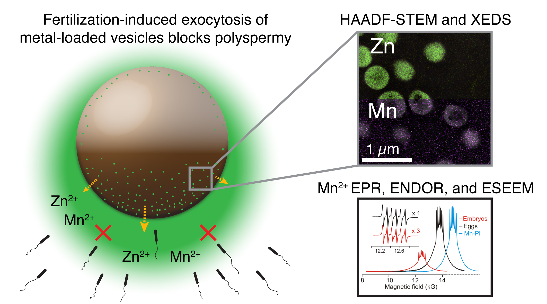
How do essential metals help an egg mature, become fertilized, and begin development? Our collaborations use mammalian, frog, and sea urchin models to investigate where metals are stored, how they are released during fertilization, and how these changes support early development.
Working with Brian Hoffman, Francesca Duncan, and Teresa Woodruff, we combine elemental imaging, microscopy, and spectroscopy to study both metal distributions and their chemical environments within eggs and embryos. These biological questions guide the development of methods that connect changes in elemental organization with the transition from egg to embryo.
Metal release during fertilization in Xenopus. Elemental imaging and spectroscopy reveal zinc and manganese stored in cortical granules and released following fertilization, supporting a role in limiting fertilization by additional sperm. Findings from Seeler et al., Nature Chemistry (2021).
How does the spatial organization of essential metals relate to brain function? Our collaborations investigate elemental distributions across brain regions and tissue structures, seeking to understand how metal regulation relates to neuronal activity and neurological disease.
These studies guide the development of sample preparation and imaging workflows that preserve elemental distributions while resolving relevant anatomical features. Combining elemental maps with complementary microscopy helps researchers connect changes in metal abundance with the organization of brain tissue.
Infection and immunity
How does infection reshape the distribution of essential metals within tissues? Our collaborations investigate the relationship between metal availability, invading pathogens, and the host response, including studies of kidney tissue infected with the fungus Candida albicans.
These questions require imaging across multiple scales, from whole-tissue patterns to individual sites of infection. Combining elemental imaging with microscopy that identifies pathogens and tissue structure helps researchers examine how local elemental environments change during infection.
Vascular implants and tissue response
Where do metals released by biodegradable vascular implants accumulate as the material breaks down? Our collaboration with Roger Guillory II investigates the distribution of implant-derived elements within surrounding vascular tissue, connecting material degradation with the local biological environment.
These samples present a measurement challenge: high metal concentrations near an implant can occur alongside trace concentrations farther into the tissue. The project guides the development of imaging and analysis methods that can capture this wide concentration range and relate elemental distributions to tissue structure.
Gut metabolism
How is copper distribution connected to metabolic function in the gut? Our collaboration investigates how disruption of sulfide metabolism affects copper abundance and localization in the colon, using models with altered sulfide quinone oxidoreductase (SQOR) activity.
This work connects broad tissue surveys by laser ablation imaging with higher-resolution synchrotron X-ray fluorescence microscopy. Aligning copper maps with tissue autofluorescence helps researchers locate elemental changes within the architecture of intestinal crypts and guides methods for combining measurements across spatial scales.
Autoimmunity and reproductive health
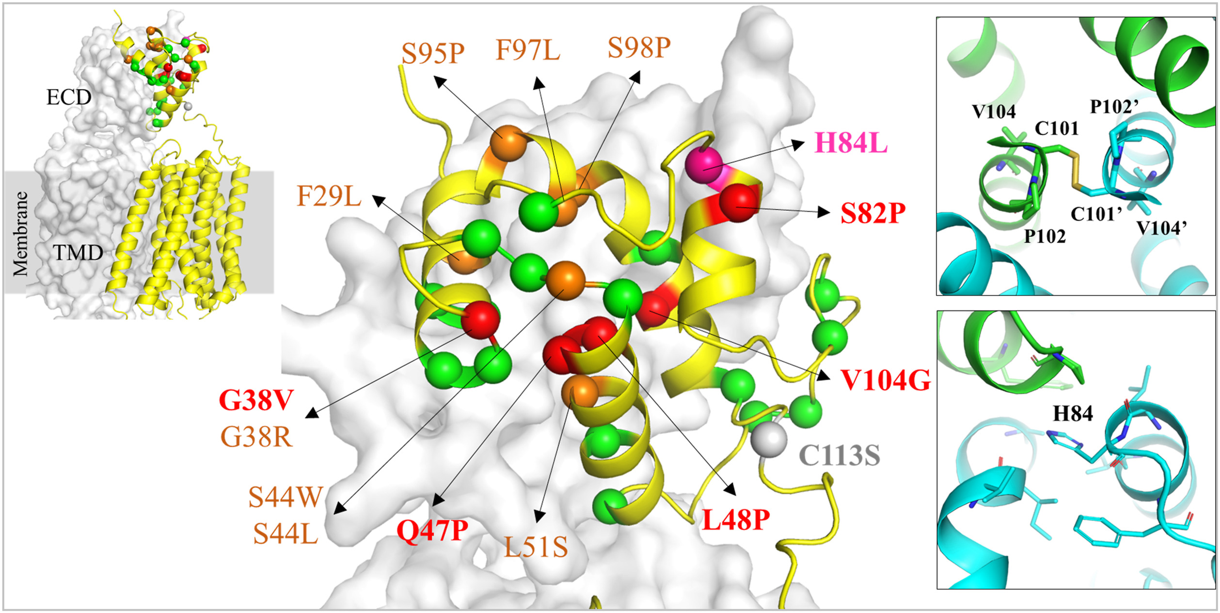
How do genetic variants change the ability of proteins to transport essential metals? Our collaboration with the Hu laboratory combines cell-based assays and quantitative elemental measurements to investigate how naturally occurring variants affect the function of the human metal transporter ZIP8.
Elemental imaging reveals where iron accumulates within the tissue. Combining these maps with anatomical and immune-cell markers helps distinguish the affected tissue compartments. This work guides improvements in spatial resolution and methods for connecting elemental distributions with specific cell populations.
How do genetic variants change the ability of proteins to transport essential metals? Our collaboration with the Hu laboratory combines cell-based assays and quantitative elemental measurements to investigate how naturally occurring variants affect the function of the human metal transporter ZIP8.
Naturally occurring variants mapped onto an AlphaFold 3 model of human ZIP8. Colored spheres locate variants studied experimentally; enlarged views highlight regions of the extracellular domain. Nikolovski et al., Journal of Biological Chemistry (2026), Figure 5.
Have a biological question that could benefit from quantitative elemental imaging? Get in touch to discuss potential research collaborations, training, or sample analysis.